
Home Industry Healthcare How 3D Medical Imaging Is Tran...
CIO Bulletin
,
10 November, 2026
Author:
CIO Bulletin Team
Lung cancer surgery is a process that requires precise decisions. Doctors need to cut enough affected tissue while protecting as much healthy lung function as possible. Achieving that is harder with earlier detection methods, particularly when tumors are buried deep inside the lungs. Below are the top ways 3D medical imaging is changing the game by giving surgeons more confidence in the operating room.
Surgeons have relied on flat 2D CT images for years to plan lung operations. The problem is that a flat image forces the operation teams to imagine what the lung actually looks like in real life. That mental reconstruction leaves room for error, particularly in complicated surgical cases.
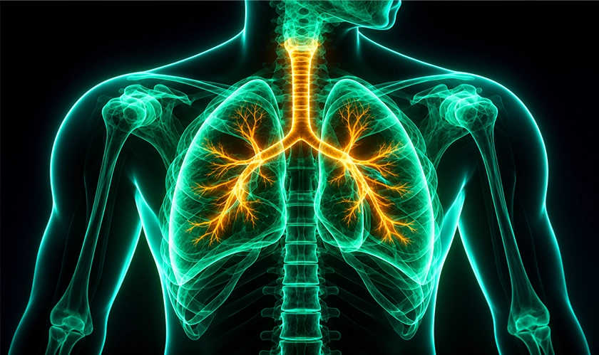
The new 3D reconstruction tools take those same CT scans and build interactive models the whole surgical team can explore. They can rotate the lung and zoom into the tumor site. This enables the team to examine exactly how the mass relates to or affects the nearby airways and blood vessels. That precision is especially useful for segmentectomies where doctors remove only a specific section of the lung, not an entire lobe.
A practical example involves surgeons at Emory Saint Joseph’s Hospital who began using this technology. They noted that it allowed them to build a clear action plan before anyone entered the operating room. That led to smoother and faster procedures.
Not every lung tumor is easy to find. Small nodules that are less than a centimeter across or located deep within the lung cannot be seen or felt during minimally invasive surgery. Surgeons working through small incisions using a thoracoscope also do not have the same feedback like those in open surgery.
Image-guided localization solves this problem. Before any operation, a radiologist uses CT imaging to mark the nodule’s exact position using techniques like hook-wire placement or injected liquid material. That marking takes the surgeon directly to the tumor during the procedure. This reduces the chance of unnecessary tissue removal or failed resection. Several studies show how such localization methods push VATS success rates to between 96% and 99% for small or hard-to-reach nodules.
3D-printed physical models of a patient’s specific lung anatomy take this a step further. Surgeons can study the images and discuss the possible approach with the patient before the first cut. This gives patients financial and legal considerations relative to their condition. For example, they can explore resources like https://www.lungcancergroup.com/legal/lung-cancer-lawyer/ to understand potential compensation claims that can help them cover medical expenses following the surgery.
One area where 3D imaging makes a difference is surgical training. 3D-printed lung models made from a patient’s CT scan give residents a chance to practice procedures on something that looks very much like real tissue. Surgical residents can learn ways to cut and stitch with staples and become comfortable with the anatomy before they step into a real operating room.
A 2025 study found that residents who trained on 3D-printed lung models scored significantly higher after training than those who only studied digital images. That is mainly because physical models gave them a hands-on understanding of the anatomy that a screen simply cannot replicate. Virtual reality simulation adds another layer. Trainees can rehearse entire procedures in a virtual environment and test different approaches to potential complications without any patient risk. This shortens their learning curve.
3D medical imaging technology supports better decisions at every stage of lung cancer surgery and helps surgeons answer key questions before the first cut. However, it does not eliminate the medical team’s responsibility to the patient. As 3D imaging tools continue to evolve, surgeons also need to acquire relevant skills to realize the technology’s value during surgery.




















































 consultants ltd.jpg)


Comments